

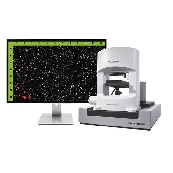
3D Cell Explorer 96focus DFT / FTC


Description
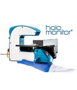
The 3D Cell Explorer 96focus is an automated live cell imaging platform designed for high-content experiments in 96-well plates. It combines label-free holotomography, three-channel epifluorescence and automated acquisition to monitor multiple cell populations over periods ranging from hours to days.
The system generates high-content, multiplexed live cell data while reducing the need for separate experiments, fluorescent reagents and manual intervention. Its automated stage, laser-based autofocus and stage-top incubation support stable, long-term observation under controlled physiological conditions.
Together with Nanolive’s AI-powered digital assays, the acquired data can be analysed and re-analysed to investigate different biological parameters without repeating the experiment.
Related products
Product description
The 3D Cell Explorer 96focus is an automated live cell imaging platform designed for high-content experiments in 96-well plates. It combines label-free holotomography, three-channel epifluorescence and automated acquisition to monitor multiple cell populations over periods ranging from hours to days. The system generates high-content, multiplexed live cell data while reducing the need for separate experiments, fluorescent reagents and manual intervention. Its automated stage, laser-based autofocus and stage-top incubation support stable, long-term observation under controlled physiological conditions. Together with Nanolive’s AI-powered digital assays, the acquired data can be analysed and re-analysed to investigate different biological parameters without repeating the experiment.Features
- Automated label-free imaging and analysis in 96-well plates
- Combination of 3D holotomography and three-channel epifluorescence
- DFT configuration: DAPI, FITC and TRITC, with optional Cy5 instead of TRITC
- FTC configuration: FITC, TRITC and Cy5
- Automated 3D and 4D time-lapse acquisition
- Resolution of 200 nm in X–Y and 400 nm in Z for holotomography
- Approximate X–Y resolution of 400 nm for fluorescence imaging
- Single field of view of 90 × 90 × 30 µm for refractive index imaging
- Extended imaging area of up to 900 × 900 × 30 µm
- Automated stage for multi-position and multi-well acquisition
- High-precision laser-based autofocus for stable long-term imaging
- Stage-top incubation for maintaining physiological conditions
- Long-term monitoring without fixation, phototoxicity or cell damage
- High-content, multiplexed live cell data
- AI-powered digital assays for automated analysis
- Retroactive analysis of previously acquired label-free data
- Walk-away acquisition with reduced hands-on time