


Showing 1–16 of 42 results
-
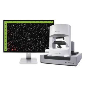
3D Cell Explorer 96focus DFT / FTC
-
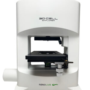
3D Cell Explorer-fluo DFT / FTC
-
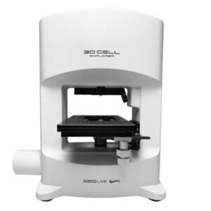
3D Cell Explorer
-

HoloMonitor – Live Cell Imaging System
-
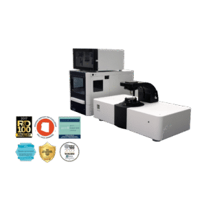
SPRm200 – Surface Plasmon Resonance Microscopy (SPRM) system
-

HoloDry
$600.00 Add to cart -

HoloLid™ for 24-Well Plate (ibidi)
$500.00 Add to cart -

HoloLid™ for 24-Well Plate (Sarstedt)
$500.00 Add to cart -
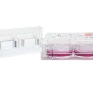
HoloLid™ for 6-Well Plate (Sarstedt)
$270.00 Add to cart -
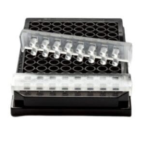
HoloLid™ for 96-Well Plate (Sarstedt)
$500.00 Add to cart -
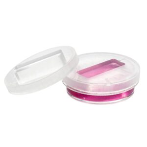
Petri Dish HoloLid™ (ibidi)
$300.00 Add to cart -
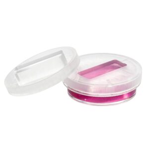
Petri Dish HoloLid™ (Sarstedt)
$300.00 Add to cart -
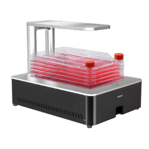
Celloger® Stack (Bright Field, 2X)
$46,253.00 Add to cart -
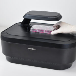
Celloger® Pro (Bright Field, Green and Red Fluorescence) with Lens Set
$74,750.00 Add to cart -
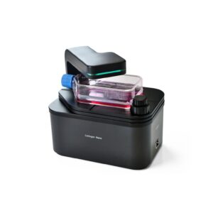
Celloger® Nano (Bright Field, 2X)
$7,130.00 Add to cart -
Celloger® Nano (Bright Field, 4X)
$7,130.00 Add to cart
Live Cell Imaging
Live cell imaging technologies enable researchers to observe and analyze living cells in real time, providing critical insights into dynamic biological processes. From cell proliferation and migration to apoptosis and drug response, live cell imaging allows continuous monitoring of cellular behavior without disrupting physiological conditions.
At Altium, we provide advanced live cell imaging and analysis solutions designed for life science research, drug discovery, cancer biology, immunology, and cell-based assay development. Our systems deliver high-resolution time-lapse imaging, automated analysis, and quantitative data generation for complex cellular studies.
What Is Live Cell Imaging?
Live cell imaging is a microscopy-based technique that allows researchers to visualize living cells over time under controlled environmental conditions. Unlike endpoint assays, this approach captures dynamic cellular processes as they happen.
Live cell imaging systems enable:
- Real-time monitoring of cell behavior
- Time-lapse microscopy acquisition
- Cell morphology and structure analysis
- Cell proliferation and viability tracking
- Drug response and toxicity studies
- 2D and 3D cell culture monitoring
By preserving cells in a controlled incubator environment, researchers can generate physiologically relevant data with minimal experimental disruption.
Advantages of Live Cell Imaging Technologies
Modern live cell imaging platforms offer significant advantages over traditional endpoint and fixed-sample microscopy:
- Continuous, non-invasive cell monitoring
- High reproducibility and experimental consistency
- Automated image acquisition and analysis
- Multi-well plate compatibility for high-throughput studies
- Long-term time-lapse experiments
- Remote access and data sharing capabilities
These capabilities are especially valuable in high-content screening (HCS) and drug discovery workflows.
Time-Lapse Microscopy and Cellular Dynamics
Time-lapse microscopy is a core feature of live cell imaging systems, enabling sequential image capture over extended periods to study dynamic biological processes.
With time-lapse imaging, researchers can:
- Track cell division and mitosis in real time
- Monitor cell migration and wound healing assays
- Observe 3D spheroid and organoid development
- Evaluate drug-induced cellular responses
- Analyze long-term phenotypic changes
This approach provides deeper biological insights compared to static imaging methods.
Fluorescence and Brightfield Imaging
Advanced live cell imaging systems support both brightfield and fluorescence imaging modalities, enabling multi-dimensional analysis of cellular processes.
These imaging modes allow researchers to:
- Visualize cellular structures in high detail
- Track specific proteins using fluorescent markers
- Perform multi-channel imaging experiments
- Study molecular signaling pathways in living cells
This combination is essential for functional cell biology and mechanistic studies.
Applications of Live Cell Imaging
Live cell imaging is widely used across biomedical and pharmaceutical research fields, including:
- Cancer biology and tumor progression studies
- Immunology and immune cell interaction analysis
- Drug discovery and toxicity screening
- Neuroscience research
- Stem cell and regenerative medicine
- Infectious disease and microbiology research
- 3D cell culture and organoid models
These applications provide physiologically relevant insights into cellular function and disease mechanisms.
Automation and High-Content Analysis
Modern live cell imaging platforms integrate automation and AI-powered analytics to improve efficiency and data quality.
Key automation features include:
- Automated focus and image acquisition
- Multi-position and multi-well scanning
- Incubator-based continuous monitoring
- AI-driven cell segmentation and classification
- Real-time data processing and reporting
These features significantly reduce manual workload while increasing experimental throughput.
Quantitative Live Cell Analysis
Beyond imaging, live cell imaging systems provide powerful quantitative analysis tools to extract meaningful biological data.
Common analysis workflows include:
- Cell counting and proliferation measurement
- Migration and invasion rate analysis
- Fluorescence intensity quantification
- Kinetic response modeling
- 3D structure analysis (spheroids and organoids)
This enables researchers to convert visual data into actionable biological insights.
Choosing the Right Live Cell Imaging System
Selecting the appropriate system depends on experimental requirements and research goals.
Key considerations include:
- Imaging resolution and sensitivity
- Fluorescence channel capabilities
- Incubator compatibility and environmental control
- Level of automation and throughput
- 2D vs. 3D imaging support
- Software and AI integration
Choosing the right platform ensures reliable data quality and experimental reproducibility.
Explore Live Cell Imaging & Analysis Solutions
Discover Altium’s live cell imaging and analysis solutions designed for real-time cellular observation, high-content screening, and quantitative biological analysis. Our technologies empower researchers to study complex cellular dynamics, accelerate drug discovery, and gain deeper insights into living systems.