

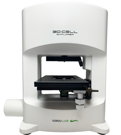
3D Cell Explorer-fluo DFT / FTC


Description
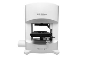
The 3D Cell Explorer-fluo is a multimodal live cell imaging platform that combines Nanolive’s label-free holotomography with three-channel epifluorescence. This combination provides detailed information about cell morphology and organelle dynamics while allowing researchers to identify specific fluorescently labelled targets within the same experiment.
Label-free imaging reduces the need for fluorescent markers and limits the effects of photobleaching and phototoxicity during long-term studies. Fluorescence can then be used selectively to visualize structures that cannot be detected through refractive index alone, including specific proteins, ions and viruses.
The system is available in DFT and FTC configurations, offering different fluorescence channel combinations to match individual experimental requirements.
Related products

Product description
The 3D Cell Explorer-fluo is a multimodal live cell imaging platform that combines Nanolive’s label-free holotomography with three-channel epifluorescence. This combination provides detailed information about cell morphology and organelle dynamics while allowing researchers to identify specific fluorescently labelled targets within the same experiment. Label-free imaging reduces the need for fluorescent markers and limits the effects of photobleaching and phototoxicity during long-term studies. Fluorescence can then be used selectively to visualize structures that cannot be detected through refractive index alone, including specific proteins, ions and viruses. The system is available in DFT and FTC configurations, offering different fluorescence channel combinations to match individual experimental requirements.Features
- Combination of label-free holotomography and epifluorescence
- High-resolution 3D and 4D live cell imaging
- Three fluorescence channels
- DFT configuration: DAPI, FITC and TRITC, with optional Cy5 instead of TRITC
- FTC configuration: FITC, TRITC and Cy5
- Label-free field of view of 90 × 90 × 30 µm
- Fluorescence field of view of 90 × 90 µm
- Correlation of cellular morphology with specific fluorescent markers
- Reduced dependence on labels during long-term experiments
- Lower photobleaching and phototoxicity compared with fluorescence-only imaging
- Visualization of proteins, ions, viruses and other specifically labelled targets
- Simultaneous observation of several cellular structures and biological processes
- STEVE software for acquisition, visualization, digital staining and data processing
- Compatibility with EVE Analytics and application-specific digital assays